A complex ball-and-socket synovial joint that connects the scapula (shoulder blade) and humerus (upper arm bone), allowing for a wide range of motion and flexibility. Also known as the glenohumeral joint, it’s a vital joint for movement and stability in the human body.
Anatomical Structure
Articulating Surfaces
The shoulder joint, also known as the glenohumeral joint, is formed by the connection between the humerus head and the scapula’s glenoid cavity. This joint features hyaline cartilage-covered articulating surfaces, typical of synovial joints. The humerus head is substantially larger than the glenoid fossa, providing a wide range of motion. To address this size disparity, the glenoid fossa is reinforced by a fibrocartilage rim called the glenoid labrum, which deepens the socket and enhances joint stability.

Reference: Shoulder Joint
Ligaments
The bear joint is upheld by a few ligaments:
– Glenohumeral Ligaments:
- Superior: connects humerus to glenoid fossa, stabilizes anterior joint aspect
- Middle: connects humerus to glenoid fossa, stabilizes anterior joint aspect
- Inferior: connects humerus to glenoid fossa, stabilizes anterior joint aspect
– Coracohumeral Ligament:
- Connects base of coracoid process to greater tubercle of humerus
- Supports superior part of joint capsule
– Transverse Humeral Ligament:
- Spans between two tubercles of humerus
- Secures the tendon of the long head of the biceps muscle within the intertubercular sulcus, preventing its displacement.
– Coracoacromial Ligament:
- Links the acromion and coracoid process, forming a bridge between these two bony projections on the scapula.
- Forms coracoacromial arch, resists superior displacement of humeral head.

Reference: Ligaments
Bursae
A bursa is a fluid-filled sac that cushions the movement of tendons, bones, and skin, minimizing friction and enabling smooth motion. In the shoulder joint, several bursae play a crucial role:
– The sub acromial bursa, nestled between the deltoid muscle and acromion, and above the supraspinatus tendon and joint capsule, facilitates frictionless movement of the rotator cuff tendons by reducing friction beneath the deltoid.
– The subscapular bursa, arranged between the subscapularis ligament and the scapula, guarantees consistent floating of the ligament amid bear developments.

Reference: Bursa Of Shoulder Joint
Movements
The shoulder joint boasts exceptional mobility, allowing for a broad range of movements:
– Extension (posterior movement of the upper limb): The posterior deltoid, latissimus dorsi, and teres major muscles work together to move the arm backward.
– Flexion (anterior movement of the upper limb): The pectoralis major, anterior deltoid and coracobrachialis muscles collaborate to move the arm forward, with the biceps brachii providing weak assistance.
– Abduction (lateral movement of the upper limb): The supraspinatus initiates the first 15 degrees of movement, followed by the middle deltoid fibers, which take over until 90 degrees. Beyond 90 degrees, scapular rotation is necessary, facilitated by the trapezius and serratus anterior.
– Adduction (medial movement of the upper limb): The pectoralis major, latissimus dorsi, and teres major muscles combine to move the arm toward the midline.
– Internal rotation (inward rotation, with the thumb pointing medially): The subscapularis, pectoralis major, latissimus dorsi, teres major and anterior deltoid work together to achieve this movement.
– External rotation (outward rotation, with the thumb pointing laterally): The infraspinatus and teres minor muscles collaborate to rotate the arm outward.
– Circumduction (circular movement of the upper limb): This movement results from a combination of the aforementioned actions.
Blood Supply
The shoulder joint’s blood supply is ensured by a trio of key arteries:
– The axillary artery’s anterior and posterior circumflex humeral branches, which encircle the joint.
– The suprascapular artery, originating from the thyrocervical trunk, which supplements the joint’s vascularization.
Innervation
The shoulder joint’s sensory innervation is provided by the axillary and suprascapular nerves.
Common Injuries
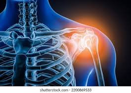
Dislocation of the Shoulder Joint:
Shoulder dislocations occur when the humeral head is displaced from the glenoid fossa. The most common type is anterior dislocation, accounting for 95% of cases, which results from excessive extension and lateral rotation. This forces the humeral head anteriorly and inferiorly, potentially leading to tearing of the joint capsule, Hill-Sachs lesions, and Bankart lesions. The axillary nerve, running near to the bear joint, is powerless to harm amid disengagement or endeavored lessening, causing deltoid loss of motion and misfortune of sensation over the regimental identification region.
Rotator Cuff Tendonitis:
Rotator cuff injuries are common due to the heavy strain on these muscles, which play a crucial role in stabilizing the glenohumeral joint. The spectrum of rotator cuff pathology includes tendinitis, shoulder impingement, and sub-acromial bursitis. Supraspinatus tendinitis is characterized by a “painful arc” during abduction indicating potential degenerative changes in the subacromial bursa and supraspinatus tendon. This may also suggest a partial tear of the supraspinatus tendon. Overuse often leads to inflammation of the muscle tendons, causing tendinitis, which can progress to more severe conditions if left untreated.

Reference: Cause of Shoulder Pain
The shoulder joint is a complex and vital part of our musculoskeletal system, playing crucial role in our daily lives. By understanding its anatomy, function, and common issues, we can take steps to prevent injuries, and ensure mobility and strength. Remember to prioritize shoulder joint care through regular exercise, proper posture, and timely treatment.